FIG. 2.
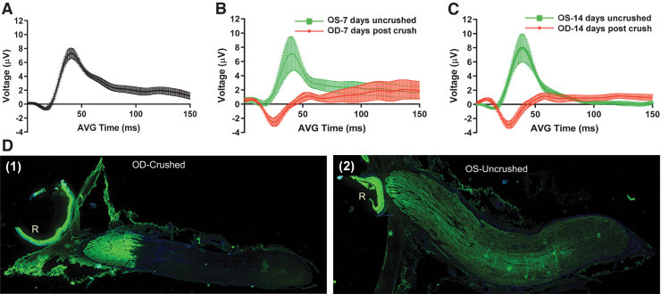
Assessment of porcine optic nerve form and function before and after optic nerve crush. (A) Baseline flash visual evoked potential (fVEP) recordings were obtained from 15 Yucatan minipigs 3–7 days prior to optic nerve crush. For all fVEP recordings, replicates were performed for oculus dextrus (OD) followed by oculus sinister (OS) (n = 30 waveforms). fVEP recording 7 days (B) and 14 days (C) after optic nerve crush from saline-only injected minipigs (n = 4 pigs, 8 waveforms per eye/per time point). (D) Axonal transport of cholera toxin-β subunit (CT-β) in uncrushed and crushed optic nerves. Two days before euthanasia and harvest of eyes and optic nerves, 500 μg of CT-β was dissolved in 250 μL of injectable saline and 100 μL of CT-β solution was injected into the vitreous of both eyes. Two days after injection, the pigs were euthanized via transcardial perfusion fixation with 10% formalin, and the eyes and optic nerves were harvested. The optic nerves and peripapillary retina were then prepared for frozen sectioning. The right (crushed) optic nerve (D1) shows fluorescent signal from the CT-β up to, but not beyond, the crush site. In the immunofluorescence pattern obtained from the left eye (D2), the CT-β signal can be seen through the entire length of the nerve, demonstrating intact, unimpaired axonal transport throughout its entire length.

