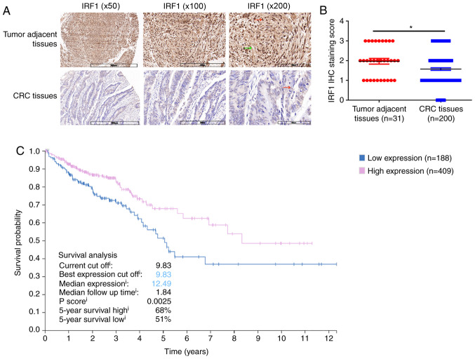
Figure 1.
IRF1 expression in CRC tissues is significantly lower than that in adjacent tissues. (A) Representative images of the different staining patterns. Scale bar, 300, 200 60 µm. Red arrows indicate the nucleus and green arrows indicate the cytoplasm. (B) IRF1 staining scores as assessed by IHC in CRC tumors (n=200) and adjacent tissues (n=31). *P<0.05. (C) Association between IRF1 expression and the 5-year survival rate. Data were acquired from the Human Protein Atlas database. CRC, colorectal cancer; IHC, immunohistochemistry; IRF1, interferon regulatory factor 1.

