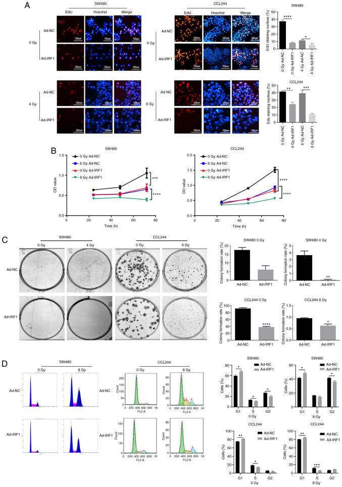
Figure 3.
Overexpression of IRF1 inhibits the proliferation of CRC cells and increases their sensitivity to X-ray irradiation. (A) Subsequently, 0 and 4 or 6 Gy X-rays were administered to the cells at 48 h after infection. At 48 h after irradiation, EdU was used to detect the proliferation of CRC cells. Scale bar, 100 or 200 µm. (B) Subsequently, 0 and 6 Gy X-rays were administered to the cells at 48 h after infection. At 24, 48 and 72 h after irradiation, MTT was used to detect the proliferation of CRC cells. (C) After 14 days, the cells were fixed with pure methanol and then stained with Giemsa crystal violet, and the cell colony formation rate was calculated. (D) After 48 h, a flow cytometer system was used to analyze the cell cycle *P<0.05, **P<0.01, ***P<0.001 and ****P<0.0001. Ad, adenovirus expression vector; CRC, colorectal cancer; EdU, 5-ethynyl-20-deoxyuridine; IRF1, interferon regulatory factor 1; NC, negative control; OD, optical density.

