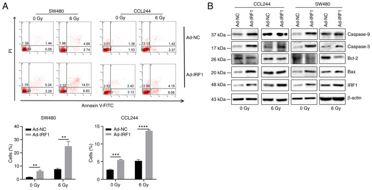
Figure 4.
Overexpression of IRF1 increases X-ray-induced apoptosis. (A) At 48 h after infection, 0 and 6 Gy X-rays were administered to the cells. After 48 h, a flow cytometer system was used to detect apoptosis of the cells. (B) At 48 h after infection, 0 and 6 Gy X-rays were administered to the cells. After 48 h, the cells were harvested for protein isolation and western blotting to detect the expression levels of the apoptosis-related proteins Bax, Bcl-2, caspase-3 and caspase-9. **P<0.01, ***P<0.001 and ****P<0.0001. Ad, adenovirus expression vector; IRF1, interferon regulatory factor 1; NC, negative control.

