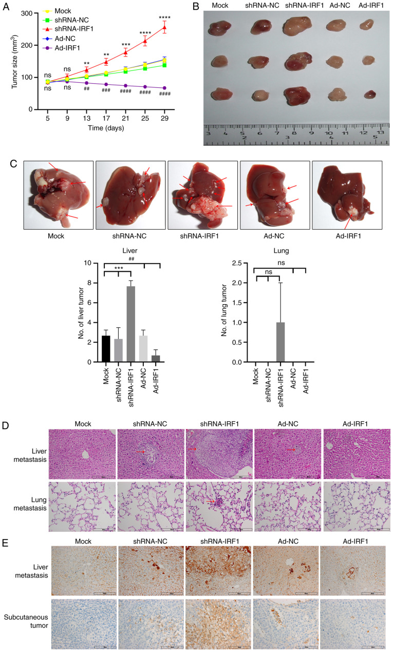
Figure 5.
Overexpression of IRF1 promotes radiosensitivity of CRC to X-rays in vivo. (A) Tumor growth curve of each group. (B) Representative tumors from each group of mice. (C) Representative CRC metastases in the liver. Red arrows indicate lesions. Analysis of the number of liver and lung metastases in each group. (D) H&E staining of nodules in the liver and lung for CRC metastasis for each group. Red arrows indicate lesions. Scale bar, 60 µm. (E) Immunohistochemical detection of Bcl-2 expression in liver metastases and subcutaneous tumors for each group. Scale bar, 60 µm. **P<0.01, ***P<0.001 and ****P<0.0001 (shRNA-IRF1 vs. shRNA-NC/mock); ##P<0.01, ###P<0.001 and ####P<0.0001 (Ad-IRF1 vs. Ad-NC/mock). Ad, adenovirus expression vector; CRC, colorectal cancer; IRF1, interferon regulatory factor 1; NC, negative control; ns, not significant; shRNA, short hairpin RNA.

