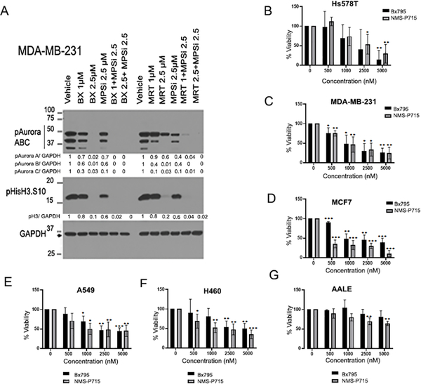
Figure 1: Inhibition of TBK1 and TTK reduces cell proliferation and viability.
(A). MDA-MB-231 cells were treated with indicated doses of TBK1 inhibitor BX795 and TTK inhibitor NMS-P715 alone or in combination for 24 hours. Western blot shows reduced levels of mitotic markers phospho-Aurora kinase and phospho-histone H3-S10, which indicates inhibition of cell proliferation. Normalized quantification for respective proteins is shown below each lane. B-F. MTT assays were performed after treatment with increasing concentrations of TBK1 inhibitor (BX795) or TTK inhibitor (NMS-P715) for 96 hours in the following breast cancer cell lines: HS578T (B); MDA-MB-231 (C); MCF7 (D); and lung cancer cell lines A549 (E); H460 (F); and Immortalized primary lung epithelial cell line AALE (G). Plots are an average of three independent experiments. Error bars represent mean ± S.D (*p<0.05, **p<0.01, ***p<0.001; one-way ANOVA).

