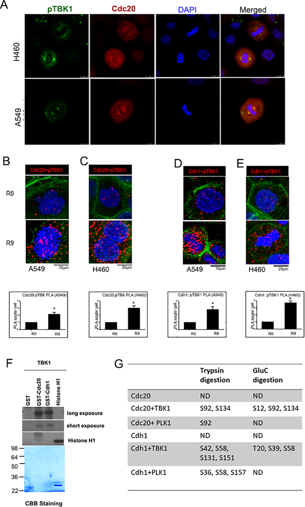
Figure 2: TBK1 interacts with and phosphorylates Cdc20 and Cdh1.
(A) Double immunofluorescence for pTBK1 (green) and cdc20 (red) was performed in A549 and H460 cells. Confocal images show a colocalization of pTBK1 with Cdc20 in the cell. (B-C). Proximity ligation assay (PLA) performed on (B) A549 and (C) H460 cells shows interaction of pTBK1 and Cdc20 in cells released from G1/ S block. D-E. PLA performed on (D) A549 and (E) H460 cells shows interaction of pTBK1 and Cdh1 in cells released from G1/ S block. Red dots represent foci of interaction of pTBK1 with Cdc20 or Cdh1, DAPI is shown in blue and phalloidin in green. Plots are an average of two independent experiments. Error bars represent mean ± S.D. (*p<0.05; unpaired two tailed t-test). (F) In vitro kinase assay shows phosphorylation of Cdc20 and Cdh1 by TBK1. (G) Phosphorylation sites were identified using mass spectrometric analysis after trypsin or GluC protease digestions.

