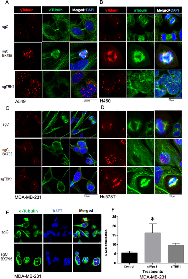
Figure 6: TBK1 inhibition and depletion results in mitotic aberrations.
TBK1 was either chemically inhibited or depleted using CRISPR/ Cas9 techniques in A549 (A), H460 (B), MDA-MB-231 (C) and Hs578T (D) cells; they were then stained for α tubulin (green) and γ tubulin (red) to study mitotic aberrations, specifically centrosomal amplification. (E) MDA-MB-231 cells were treated with 2.5μM BX795 for TBK1 inhibition and stained with α Tubulin and DAPI show multinucleation. (F) Micronucleation was studied in MDA-MB-231 cells after knockdown of TBK1 or TTK using siRNA transfection. A total of 200 cells were considered and micronuclei counted for each condition for quantification. Plot represents mean of three independent experiments. A one-way ANOVA-test was done for significance. p≤ 0.05 was considered significant. Error bars are mean ± S.D.

