Figure 7.
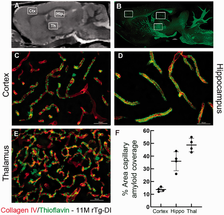
Pattern of microvascular fibrillar Aβ deposition in 11 M rTg-DI rats. (a) Sagittal plane from a proton-density-weighted MRI of a WT rat highlighting anatomical areas of interest: Cortex (Ctx), hippocampus (Hip) and thalamus (Thal). (b) Sagittal brain section from an 11 M old rTg-DI immunolabeled with thioflavin S (ThS) to identify fibrillar amyloid (green). The white boxes highlight the anatomical areas magnified in c–e which are sections from these areas now immunolabeled with rabbit polyclonal antibody to collagen IV to specifically detect cerebral microvessels (red) and stained with ThS to identify fibrillar amyloid (green). The microvascular amyloid deposits are clearly present in Ctx (c), in the hilus region of the hippocampus (d) and in the thalamus (e). Scale bars = 50 µm. (f) The quantitation of microvascular amyloid load in the three brain regions confirming the thalamic area is most heavily affected.

