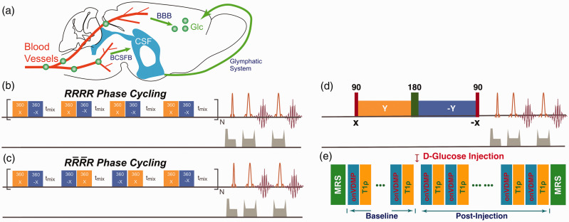
Figure 1.
(a) Illustration of the glucose transport pathways in mouse brain after intravenous infusion of glucose. Glucose in blood crosses the BBB’s luminal and abluminal membranes and reaches parenchyma. Part of the glucose rapidly enters the CSF through the BCSFB and recirculates to the parenchyma through the glymphatic system. The onVDMP saturation module with (b) and (c) phase cycling is composed of a train of bionomical pulses separated by a delay tmix. The flip angle of the binomial pulse is 360°. In the current study, a simple binomial pulse composed of two high power pulses with opposite alternating phase () was used. (d) T1ρ method: the water magnetization is first flipped by a hard 90° pulse and then locked by a spin-lock pulse. The hard 90° pulse following the spin-lock pulse flips the magnetization back to the Z-axis for MRI readout. The 180° pulse in the middle of spin-lock pulse was used to improve the robustness to the B0 inhomogeneity. (e) Diagram for the DGE study using both onVDMP and T1ρ sequences, which were collected in an interleaved manner. Two MRS spectra were collected before and after the experiment to confirm the glucose and other metabolites variation during glucose infusion in brain.
BBB: blood–brain barrier; CSF: cerebrospinal fluid; BCSFB: blood–cerebrospinal fluid barrier; MRS: magnetic resonance spectroscopy; onVDMP: on-resonance variable delay multiple pulse.

