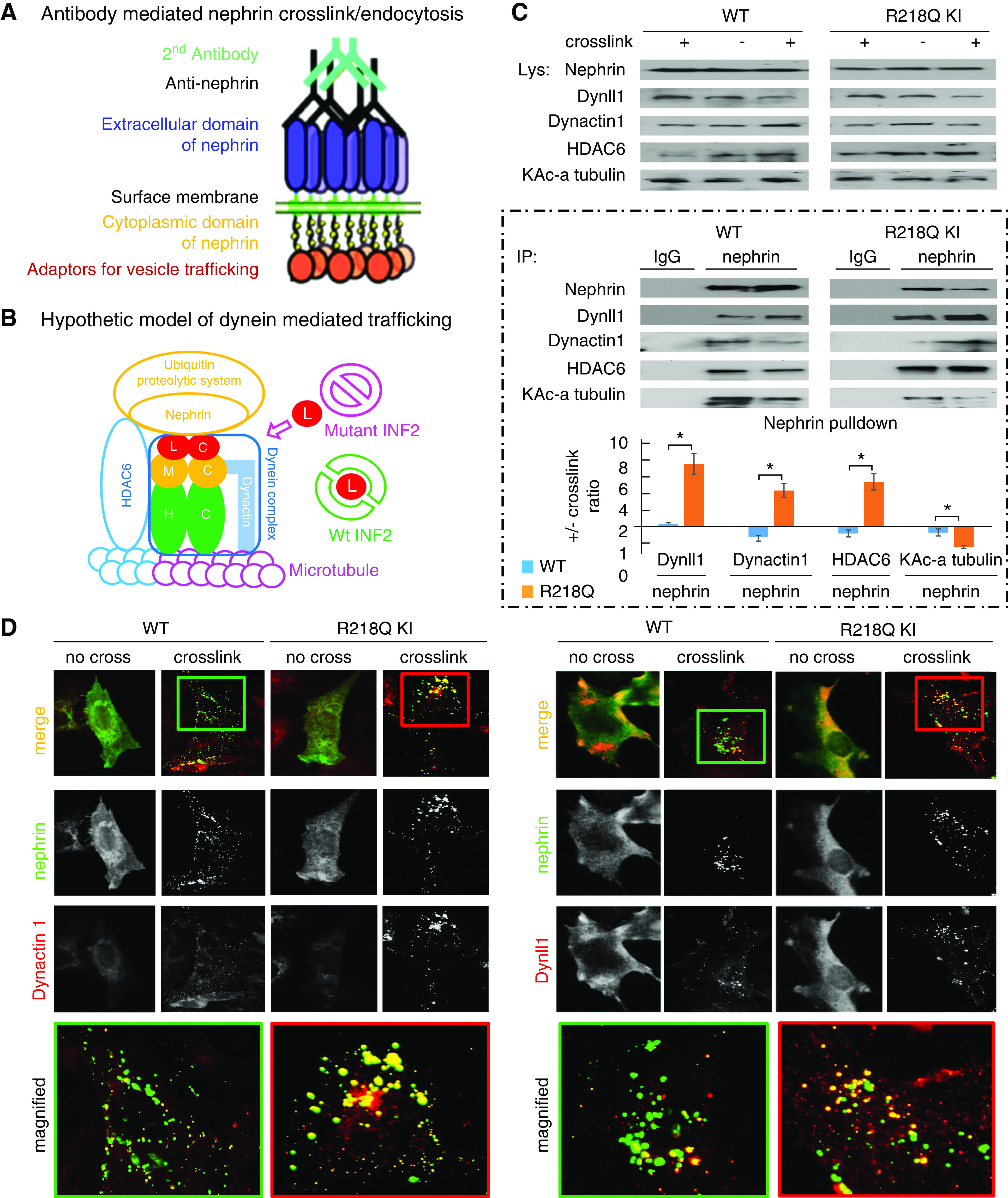
Figure 3.

Enhanced dynein involvement in postendocytic sorting of nephrin. (A) Schematic of antibody-mediated nephrin crosslink and endocytosis. Podocytes overexpressing nephrin are incubated in media containing anti-nephrin antibody followed by Alexa Fluor 488–labeled secondary antibody to induce crosslinking and endocytosis of nephrin. (B) Hypothetic schematic model of dynein-mediated nephrin trafficking along the microtubule, coupled to UPS by the bridging of HDAC6. (L, Dynll1; M, dynein intermediate chains; H, dynein heavy chains; blue tubulin, deacetylated alpha tubulin; pink tubulin, acetylated alpha tubulin). (C) WT podocytes or R218Q KI podocytes with or without antibody-mediated nephrin crosslink were lysed and subjected to IP with anti-nephrin (using IgG as a negative control). The Dynll1, Dynactin 1, HDAC6, and the Lysine acetylated alpha tubulin in nephrin pulldown were analyzed by immunoblotting, quantified in Image J, and normalized to nephrin. The ratios of these normalized levels in cells with crosslink to those without crosslink were calculated. For statistical analysis, ratios of normalized Dynactin 1, HDAC6, and the Lysine acetylated alpha tubulin in cells +/- crosslink from three independent experiments were compared using independent sample t test. *P<0.05 R218Q versus WT. (D) Immunofluorescent-based colocalization illustrated the recruitment of Dynactin 1 and Dynll1 (red) to endocytosed nephrin (green) after antibody-mediated crosslinking.
