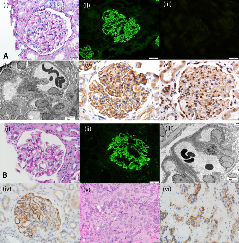
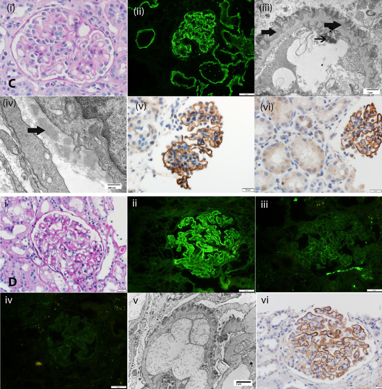
Figure 1.
Biopsy specimen findings of EXT1/EXT2-, NELL1-, Sema3B-, and PCDH7-associated MN. (A) EXT1/EXT2-associated MN: (i) Periodic acid–Schiff stain showing thickened glomerular capillary walls. Original magnification, ×40. (ii) IF microscopy showing bright C3 staining along glomerular capillary walls; (iii) IF microscopy is negative for PLA2R; (iv) electron microscopy showing subepithelial electron dense deposits. Original magnification, ×2900. IHC showing (v) EXT1 and (vi) EXT2 staining along the GBM. (B) NELL1-associated MN: (i) Periodic acid–Schiff stain showing thickened glomerular capillary walls. Original magnification, ×40. (ii) IF microscopy showing bright C3 staining along glomerular capillary walls; (iii) electron microscopy showing subepithelial electron dense deposits. Original magnification, ×2900. (iv) IHC showing NELL1 staining along the glomerular capillary walls; (v) hematoxylin and eosin stain showing squamous cell carcinoma; (vi) IHC showing that the tumor cells are positive for NELL1. (C) Sema3B-associated MN: (i) Periodic acid–Schiff stain showing thickened glomerular capillary walls. Original magnification, ×40. (ii) IF microscopy showing bright IgG staining along the GBM and TBM. Original magnification, ×20. (iii) Electron microscopy showing subepithelial electron dense deposits along GBM and tubuloreticular inclusions in endothelial cells. Original magnification, ×11,000. (iv) Electron microscopy showing TBM electron dense deposits. Original magnification, ×30,000. (v) IHC showing Sema3B staining along the GBM. Original magnification, ×40. (vi) IHC showing Sema3B staining along GBM but negative staining along the TBM. Original magnification, ×20. Arrows point to tubular basement membrane deposits. (D) PCDH7-associated MN: (i) Periodic acid–Schiff stain showing thickened glomerular capillary walls. Original magnification, ×40. (ii) IF microscopy showing bright IgG staining along the GBM. Original magnification, ×240. (iii) IF microscopy showing trace staining for C3, and (iv) negative staining for C1q. (v) Electron microscopy showing subepithelial electron dense deposits alongGBM. Original magnification, ×6800. (vi) IHC showing PCDH7 staining along the GBM. Original magnification, ×40.


