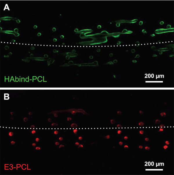
Figure 8.

Confocal fluorescence microscopy image of HAbind/E3-PCL scaffold showing peptides are presented in opposing zones. Cross-sections were labeled with (A) fluorescein-tagged HA (fl-HA; green) to bind HAbind peptides or (B) amino-Cy3 (red) to react with carboxyl groups on E3 peptides.
