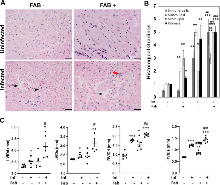
Fig 8. Loss in fat cells exacerbates cardiac pathology and causes bi-ventricular enlargement in the hearts of chronic CD mice.
A. Hematoxylin and eosin (H&E) staining of hearts in indicated mice (infected or uninfected mice, fat-ablated (Fab +) or fat-unablated (Fab-)). Infiltrated immune cells, red arrowhead; vasculitis, black arrowhead; and presence of lipid droplets, black arrow. Bar = 100 μm, 20x magnification. B. Histologic grading of heart tissue pathology was carried out according to experimental groups and classified in terms of degree of infiltrated immune cells, size of adipocytes (macro lipid and micro lipid droplets), and fibrosis in the H&E sections of hearts in chronic T. cruzi infected mice with and without fat ablation (5 images per section/mouse in each group). Each class was graded on a six-point scale ranging from 0 to 5+ as discussed in Method section and presented as a bar graph. The values plotted are mean ± standard deviation (SD) from n = 5. C. Cardiac ultrasound imaging analysis in indicated mice (infected or uninfected mice, fat-ablated (Fab +) or fat-unablated (Fab-) 90DPI). Left ventricle internal diameter (LVID) and right ventricle internal diameter (RVID) both at diastole (d) and systole (s) conditions. The error bars represent SEM. A.U. indicates arbitrary unit. *p≤0.05, **p≤0.01 or ***p≤0.001 compared with uninfected fat-unablated. #p≤0.05, ##p≤0.01 or ###p≤0.001 compared with infected fat-unablated.

