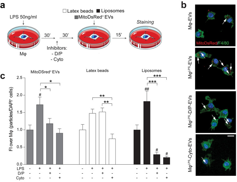
Fig 7. Pro-inflammatory mononuclear phagocytes uptake EV-associated mitochondria via endocytosis.
(a) In vitro experimental setup of EV uptake studies in MφLPS. MφLPS were treated with either Cyto or D/P and then exposed to MitoDsRed+ EVs (1:30). Latex beads and liposomes were used as positive controls of phagocytosis and endocytosis, respectively. (b, c) Representative confocal microscopy images (maximum intensity projection of Z-stacks) and quantification of MitoDsRed+ EV (red) uptake in MφLPS (stained for F4/80, green) in the presence or absence of endocytosis (D/P) and actin mediated phagocytosis/endocytosis (Cyto) inhibitors. Nuclei are stained with DAPI (blue). Data are mean FI over unstimulated Mφ (± SEM) from N ≥ 8 ROIs per condition. #p < 0.05, ##p < 0.01 vs. unstimulated Mφ. *p < 0.05, **p < 0.01, ***p < 0.001. Scale bars: 10 μm. (Data available in S3 Data). Cyto, Cytochalsin; D/P, Dynasore and Pitstop 2; DAPI, 4′,6-diamidino-2-phenylindole; EV, extracellular vesicle; FI, fold induction; LPS, lipopolysaccharide; ROI, region of interest.

