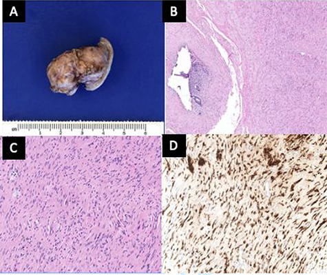
Figure 5 .

(A) The outer surface of the appendix was smooth to slightly rough with cauterized fibrous adhesions. (B) The appendiceal wall was diffusely expanded by an irregular spindle cell proliferation involving the mucosa, submucosa and muscularis propria. A tiny lumen was identified in this section, but the other sections showed completely obliterated central lumen, mimicking fibrous obliteration. (C) High-power view of spindle cells in the muscularis propria demonstrated wavy dark nuclei. (D) Spindle cells were positive for S100.
