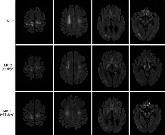
Figure. Serial Brain MRIs in Presented Case.
Diffuse leukoencephalopathy with restricted diffusion in the corona radiata and subcortical white matter on the first MRI slightly decreased on follow-up MRIs. Newly developed restricted diffusion of the globus pallidus and substantia nigra was seen on the second and third MRIs. No signs of hemorrhages, territorial infarcts, or microbleeds were seen.

