Figure 1.
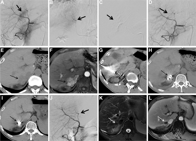
Images from case 15. Male, 54 years of age, HCC. The lesion is located at S7 with a diameter of 3.3cm.
Notes: (A) Selective angiography of the common hepatic artery during the second cTACE procedure shows tumor staining (arrow); (B and C) superselective embolization of the tumor feeding artery (arrow); (D) selective angiography of the common hepatic artery shows tumor staining disappears after embolization (arrow); (E) follow-up CT scan 2 months after thesecond cTCAE shows little lipiodol accumulation within the HCC lesion (arrow); (F) follow-up contrast-enhanced MRI 2 months after thesecond cTCAE shows persistent contrast enhancement of the HCC lesion (arrow); (G) 125Ibrachytherapy for TACE-refractory HCC is performed under the guidance of CT (arrow); (H) CT 1 month after 125Ibrachytherapy shows HCC lesion is stable (arrow); (I) CT 6 months after 125Ibrachytherapy shows remarkable particles aggregation (arrow); (J) selective angiography of the common hepatic artery 6 months after 125Ibrachytherapy shows no visible tumor staining (arrow); (Kand L) T2WI and contrast-enhanced MRI 6 months after 125Ibrachytherapy show complete response with 100% necrosis of the TACE-refractory HCC lesion (arrow).
Abbreviations: HCC, hepatocellular carcinoma; cTACE, conventional transarterial chemoembolization; CT, computed tomography; MRI, magnetic resonance imaging; T2WI, T2-weighted imaging; TACE, transarterial chemoembolization.

