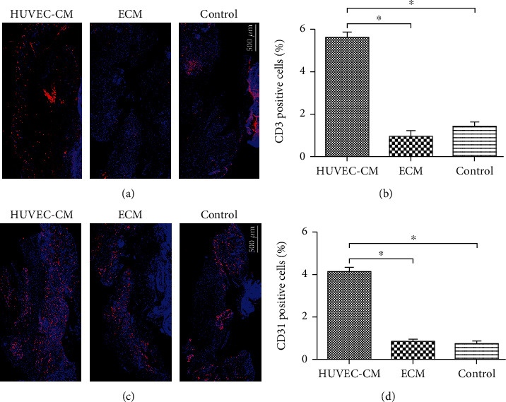
Figure 7.

Expression of lymphocyte marker CD3 and angiogenesis marker CD31 at the defect site. (a) Immunofluorescence microscopy showing endothelial cell immunostaining for CD31 (red) and measurement and analysis of CD31-positive cells. (b) Immunofluorescence microscopy showing endothelial cell immunostaining for CD3 (red) and measurement and analysis of CD3-positive cells.
