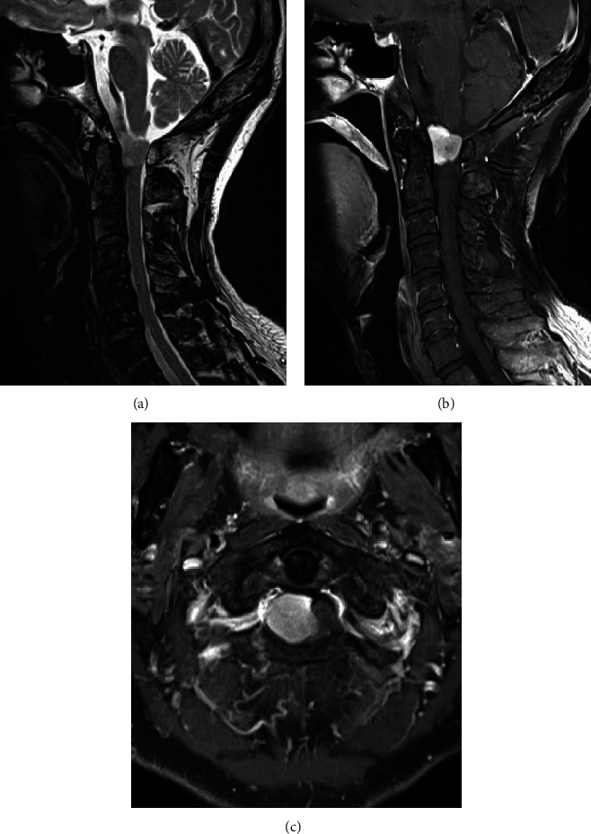
Figure 1.

(a) T2-weighted sagittal MRI of the cervical spine showed a tumor in the craniocervical junction. (b, c) MRI followed by the intravenous administration of gadolinium-diethylenetriaminepentaacetic acid (Gd-DTPA) showed a homogeneously enhanced intradural tumor without obvious dural tail sign.
