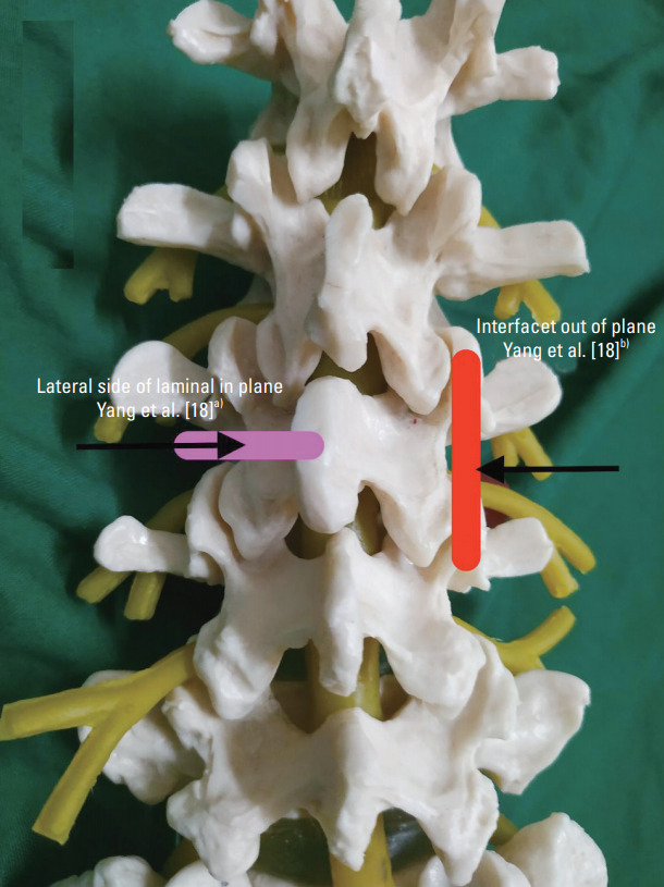
Fig. 3.

Combined approach of ultrasound transforaminal injection. Pink box, Yang et al. [18]a): First part of block. Linear probe placed over spinous process to view spinous process, lamina, and transverse process. Needle was inserted in plane, and needle tip was placed at lateral side of narrowest lamina. Red box, Yang et al. [18]b): Second part of block. Probe turned 90° to confirm needle in the middle of adjacent facets. Needle was moved out a little, slipped beside the lamina, and advanced a little inside until no resistance was felt.
