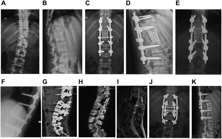
Figure 2.
Typical Case 1: A 26-year-old female patient with L1 giant cell tumor of bone. (A and B) Positive and lateral X-ray films of the lumbar spine when the patient was admitted for the first time. Pathological fracture of the lumbar vertebra, noticeable compression of the vertebral body, and spine instability were observed. (C and D) X-ray films of the positive and lateral positions of the lumbar vertebrae after tumor resection. The L1 vertebral body was resected intraoperatively, showing good positive and lateral positions of the instrumentation. (E and F) Positive and lateral X-ray films with double rods broken 32 months after operation. Broken rods occurred at the upper edge of the titanium mesh, and the titanium mesh was embedded into the upper vertebral body. (G and H) The coronal plane of lumbar CT and 3D reconstruction shows broken rods and spinal instability. (I) No tumor recurrence s found on T2-weighted MRI of the lumbar vertebrae. (J and K) The positive and lateral x-ray film after revision, fixed with double rods during the operation.

