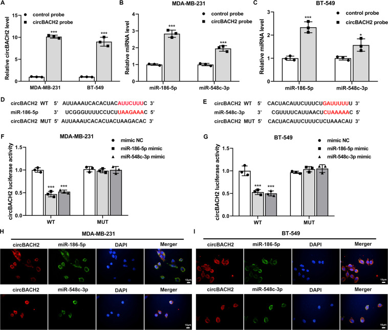
Fig. 4. circBACH2 functioned as a sponge for miR-186-5p and miR-548c-3p in TNBC cell lines.
A–C RNA pull-down assay was performed in MDA-MB-231 and BT-549 cells using a biotin-labeled circBACH2 probe, following by qRT-PCR. The control probe was used as a negative control. D, E The predicted binding sites between circBACH2 and miR-186-5p/miR-548c-3p. F, G The luciferase reporter assay was performed in MDA-MB-231 and BT-549 cells that were co-transfected with circBACH2 wild type (WT)/mutant type (MUT) luciferase vector and miR-186-5p mimic/miR-548c-3p mimic/mimic NC. H–I Fluorescence in situ hybridization (FISH) was executed to confirm the location of circBACH2 (red) and miR-186-5p/miR-548c-3p (green) in MDA-MB-231 and BT-549 cells. Nuclei were stained blue with DAPI. Scale bar = 10 µm. *P < 0.05, ***P < 0.001 vs control probe or mimic NC.

