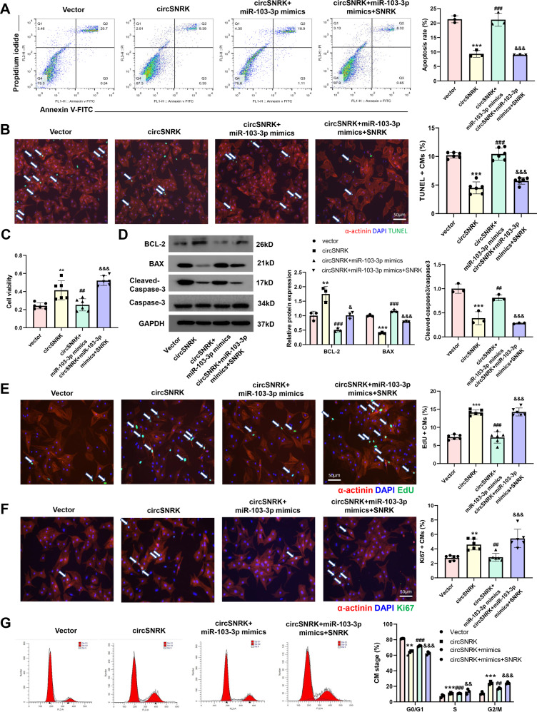
Fig. 4. The miR-103a-3p-SNRK regulatory cascade underlies cardioprotection by circSNRK.
A–C Primary cardiomyocytes were transfected with Vector, circSNRK, circSNRK + miR-103-3p mimics and circSNRK + miT-103-3p mimics + SNRK in H/SD conditions. Flow cytometry analysis (n = 3) (A) and TUNEL analysis (B) in primary cardiomyocytes after circSNRK, miR-103-3p, and SNRK interference. White arrows indicate apoptotic cells. bar = 50 μm. And Cell Counting Kit-8 (CCK-8) assay (n = 6). C **P < 0.01, ***p < 0.001 vs. Vector, ##P < 0.01, ###p < 0.001 vs. circSNRK, &&&p < 0.001 vs. circSNRK + miR-103-3p mimics (n = 6). D BCL-2, BAX, cleaved-caspase-3/caspase-3 levels in hypoxic and ischemic primary cardiomyocytes after circSNRK, miR-103-3p, and SNRK interference. **P < 0.01, ***p < 0.001 vs. Vector, ##P < 0.01, ###p < 0.001 vs. circSNRK, &P < 0.05, &&&p < 0.001 vs. circSNRK + mimics (n = 3). E EdU staining in isolated NRCMs after circSNRK, miR-103-3p, and SNRK interference and quantification of EdU positive primary cardiomyocytes. White arrows indicate EdU positive primary cardiomyocytes. ***p < 0.001 vs. Vector, ###p < 0.001 vs. circSNRK, &&&p < 0.001 vs. circSNRK + miR-103-3p mimics; bar = 50 µm. (n = 6). G Ki67 immunofluorescence staining in isolated primary cardiomyocytes after circSNRK, miR-103-3p, and SNRK interference and quantification of Ki67 positive primary cardiomyocytes. White arrows indicate Ki67-positive primary cardiomyocytes. **P < 0.01 vs. Vector, ##P < 0.01 vs. circSNRK, &&&p < 0.001 vs. circSNRK + miR-103-3p mimics; bar = 50 µm. (n = 6). H Flow cytometry analysis of primary cardiomyocytes transfected with miR-NC and miR-103-3p inhibitor. **P < 0.01, ***p < 0.001 vs. vector group, ##p < 0.01 ###p < 0.001 vs. circSNRK, &&p < 0.01 &&&p < 0.001 vs. circSNRK + miR-103-3p mimics (n = 3).

