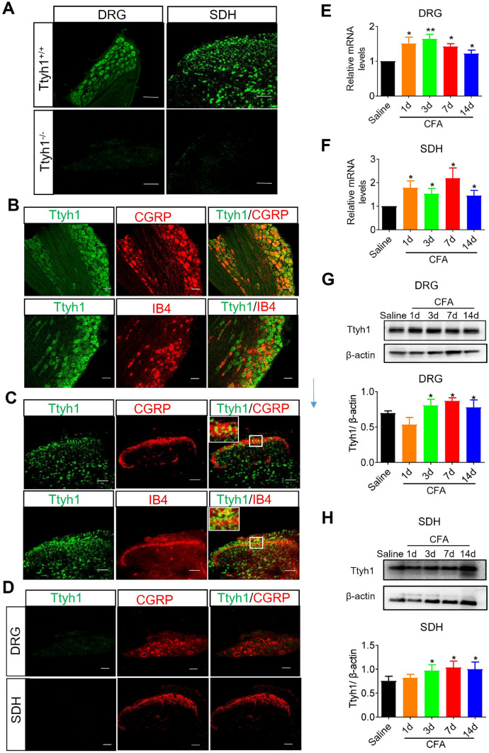
Fig. 3.
The expression and upregulation of Ttyh1 in the dorsal root ganglion (DRG) and spinal dorsal horn (SDH) following peripheral inflammation. A Fluorescence in situ hybridization of Ttyh1 (green) in DRG neurons and SDH neurons (n = 3 mice). B Double immunofluorescence staining of Ttyh1 (green) with CGRP (red, upper panels) or IB4 (red, lower panels) in the DRG (n = 6 mice). C Double immunofluorescence staining of Ttyh1 (green) with CGRP (red, upper panels) or IB4 (red, lower panels) in the SDH (n = 6 mice). D Double immunofluorescence staining of Ttyh1 (green) with CGRP (red) in the DRG (upper panels) and SDH (lower panels) in Ttyh−/− mice (n = 6 mice). E, F Upregulation of Ttyh1 at the mRNA level in the DRG (E) and SDH (F) at different time points following peripheral CFA inflammation (n = 5 mice). G, H Typical examples and quantitative summary showing upregulation of Ttyh1 at the protein level in the DRG (G) and SDH (H) at different time points following CFA injection (n = 8 mice). All data are presented as mean ± SEM. *P < 0.05, **P < 0.01. Scale bars, 50 μm.

