Fig. 6.
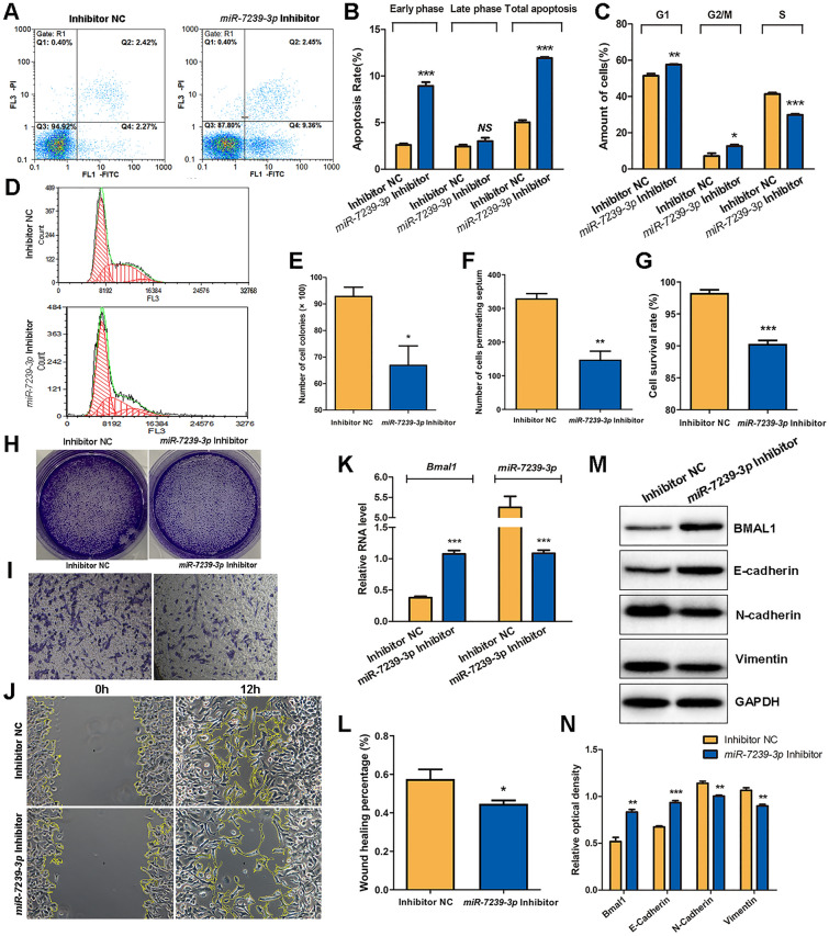
miR-7239-3p inhibitor reversed the tumor-promoting effects of M2 phenotype microglial exosomes. A Cell apoptosis and cell cycle of GL261 were detected by FCM following PI staining. The abscissa represents Annexin V, and the ordinate represents PI. B FCM results of Apoptosis. C, D FCM results of cell cycle. The abscissa of D represents the DNA content and the ordinate represents the number of effective cells. E The statistical results of the colony number in each group. F The statistical result of the number of cells penetrating the chamber in each group. G The statistical results of CCK8 assay for cell survival. H The formed clones of GL261 cells after crystal violet staining. I The microscopic image of GL261 cells penetrating the chamber after crystal violet staining (×100). J The microscopic image of GL261 cell scratch experiments. K The relative expression levels of Bmal1 and miR-7239-3p RNA in the two groups were detected by real-time qPCR. The internal reference protein is β-actin. L The statistical results of cell scratch experiments. The vertical axis represents the wound healing percentage. M Protein bands of relevant proteins in the two groups were detected by Western blot. N Quantitative analysis of protein expressions (n = 3). *P < 0.05, **P < 0.01, ***P < 0.001.

