Fig. 3.
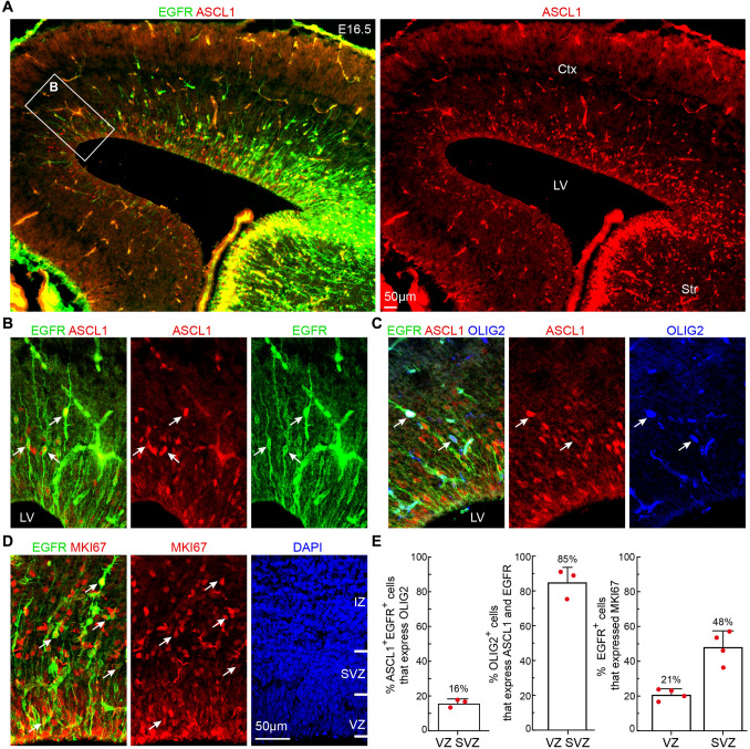
Identification of aMIPCs and bMIPCs in E16.5 cortex. A ASCL1 and EGFR expression in the cortex at E16.5. Note that both ASCL1 and EGFR are expressed in a high lateral (ventral) to low medial (dorsal) gradient in the cortex. B Higher magnification of the boxed area in A showing bipolar ASCL1+EGFR+ aMIPCs (arrows). C OLIG2 expression in ASCL1+EGFR+ cells (arrows). Non-specific binding of OLIG2 mouse monoclonal antibody to E16.5 brain sections is evident. D EGFR+MKI67+ cells in the cortex (arrows). E Quantification of the above immunostaining experiments. Mean ± SEM (n = 3–4 mice). Ctx, cortex; IZ, intermediate zone; LV, lateral ventricle; Str, striatum.

