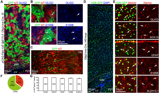
Fig. 7.
Cortical RGC-derived Olig2+ bMIPCs give rise to the vast majority of cortical oligodendrocytes and astrocytes, and a subpopulation of OBiNs. A Representative images showing GFP+ cells derived from cortical RGCs (pCAG-Flpo plasmids were electroporated to the cortical VZ of Olig2-tva-Cre; IS mice at E15.5 and the brains were analyzed at P21). B Higher magnification image showing GFP+OLIG2+ (arrow) and GFP+S100B+ cells (arrows) in the cortex. C GFP+ interneurons and tdT+ cells in the OB. D H2B-GFP+ cells in the P21 cortex of an Olig2-tva-Cre; HG loxp mouse. E Double immunofluorescence labeling for H2B-GFP and markers (arrows) for cortical oligodendrocytes (OLIG2 and SOX10) and astrocytes (GFAP, S100B, and ALDH1L1). F Percentages of GFP+S100B+ astrocytes vs GFP+OLIG2+ oligodendrocytes in the cortex (n = 3 mice). G Histogram summarizing the percentages of H2B-GFP+ cells that express different markers (n = 3 mice).

