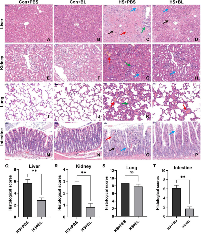
FIGURE 3.
BL pre-administration attenuated multiple-organ injury in HS rats. Organ samples were harvested at 3 h after HS and stained with H&E. Representative histopathological images are shown of liver (A–D), kidney (E–H), lung (I–L), and intestine (M–P) from Con + PBS, Con + BL, HS + PBS, and HS + BL (left to right) groups, magnification 200×. Scale bar = 50 μm. Arrows indicate typical pathological changes including necrosis, inflammation, vacuolar degeneration, congestion, hemorrhage, and alveolar wall thickening. Histological scores of liver (Q), kidney (R), lung (S), and intestine (T) were counted and plotted. Values are presented as means ± SEM; n = 6 per group. nsP > 0.05, ∗∗P < 0.01. Black arrows indicate necrosis; green arrows, inflammation; red arrows, congestion or hemorrhage; blue arrows, organ-specific alternations, such as villi destruction or hepatocyte vacuolar degeneration.

