FIGURE 4.
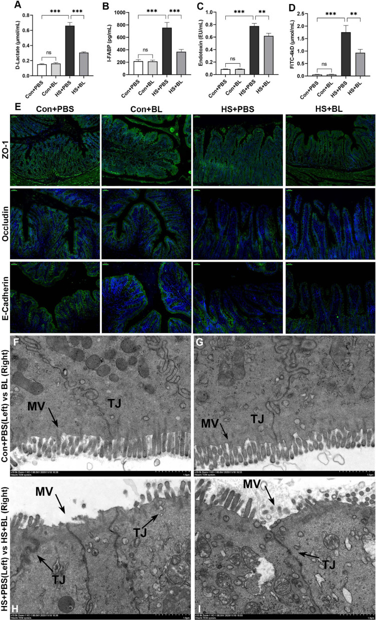
BL pre-administration attenuated intestinal injury and enhanced intestinal barrier function. Plasma D-Lactate (A), I-FABP (B), endotoxin (C), and FD4 (D) were detected at 3 h after the HS onset in the Con + PBS, Con + BL, HS + PBS, and HS + BL groups. Concentrations are presented as means ± SEM; n = 10 per group. nsP > 0.05, ∗∗P < 0.01, ∗∗∗P < 0.001. (E) Representative images of intestinal sections of rats from Con + PBS, Con + BL, HS + PBS, and HS + BL groups. Cell nuclei were stained using DAPI (blue), and ZO-1, occludin, and E-cadherin proteins were stained with corresponding antibodies (green). Scale bar = 50 μm, n = 6 per group. (F–I) Representative ultrastructural transmission electron photomicrographs of intestinal samples from Con + PBS (F), Con + BL (G), HS + PBS (H), and HS + BL (I) show the morphology and sizes of cell nuclei, membrane microvilli (MV, arrows), and tight junctions (TJ, arrows). Scale bar = 1 μm. n = 3 per group.

