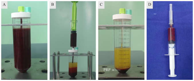
Figure 1.
Preparation of PRP. (A) Peripheral blood samples were collected from patients (50 ml each), transferred to separation tubes and then centrifuged twice at 378 x g for 10 min each. (B) Erythrocytes were removed after the first centrifugation step, and (C) the second centrifugation resulted in a middle layer of PRP, (D) which was collected using a syringe. PRP, platelet-rich plasma.

