Fig. 1.
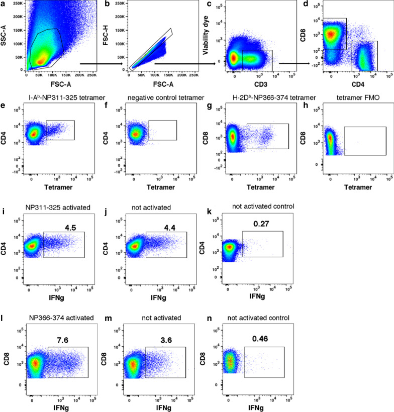
Gating of influenza antigen-specific T cells in the lung. a–d Sequential gating of lymphocytes, singlets, viable CD3 + T cells, and CD4 + and CD8 + T cells on day 7 p.i. e, f Gating of CD4 + T cells binding the I-Ab-NP311-325 tetramer or the negative control I-Ab-PVSKMRMATPLLMQA tetramer on day 7 p.i. g, h Gating of CD8 + T cells binding the H-2Db-NP366-374 tetramer or the tetramer FMO control on day 7 p.i. i, j Gating of IFN-γ + CD4 + T cells activated with the I-Ab-binding NP311-325 peptide for 5 h ex vivo or left unstimulated on day 7 p.i. Note that peptide does not augment the production of IFN-γ because the T cells remain activated in vivo at this time point. k From a poor responder mouse, gating of CD4 + T cells left unstimulated for 5 h ex vivo shows few IFN-γ + cells, which serves as a negative control for anti-IFN-γ Ab staining. l, m Gating of IFN-γ + CD8 + T cells activated with the H-2Db-binding NP366-374 peptide 5 h ex vivo or left unstimulated on day 7 p.i. n From a poor responder mouse, gating of CD8 + T cells left unstimulated for 5 h ex vivo shows few IFN-γ + cells, which serves as a negative control for anti-IFN-γ Ab staining. The examples shown are from non-smoking mice

