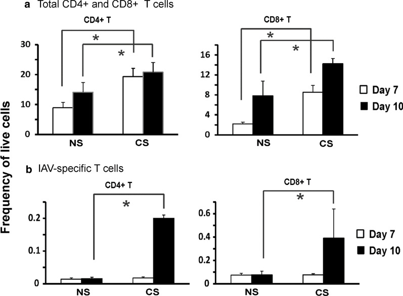
Fig. 4.
CS altered total CD4 + and CD8 + T cells and IAV-specific CD4 + and CD8 + T cells in MLN of IAV-infected mice. C57BL/6 mice were exposed to CS or not for 6 weeks in a smoke exposure chamber, then CS-exposed and NS mice were intranasally inoculated with a sublethal dose of IAV (300 pfu/mouse). At 7- and 10-days post IAV infection, MLN cells were isolated and stained for flow cytometry to determine the frequency of live cells of total CD4 + and CD8 + T cells (a). Cells from lung were stained for flow cytometry using MHC I or MHC II-tetramers containing viral NP peptides to identify IAV antigen-specific CD4 + and CD8 + T cells (b). Bar graph represents mean ± SEM (n = 5). * denotes a significant difference between CS and NS groups at the same time point

