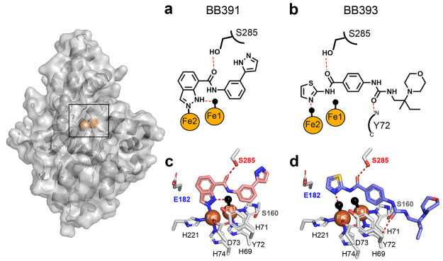
Figure 2.
Crystal structures of PqsE-BB391 and PqsE-BB393 complexes. Surface and cartoon representations of full-length PqsE shown with iron atoms (orange spheres). In a and b, key interactions for each ligand are depicted with hydrogen bonds shown as red dashed lines and direct bonds are shown as solid lines. Water molecules are depicted as black spheres. In c and d, side chains of select amino acids, including the 69HXHXDH74 motif, that form the PqsE active site are shown in gray with nitrogen atoms in blue and oxygen atoms in red. BB391 and BB393 nitrogen and oxygen atoms use the same coloring scheme. Carbon atoms are shown in pink for BB391 and light blue for BB393, and the sulfur atom in BB393 is in yellow. Hydrogen bonds, water molecules, and iron atoms appear as in a and b.

