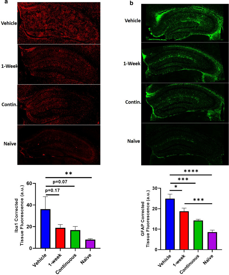
Fig 6.
CR2Crry treatment decreases astrocytosis in the contralateral hippocampus. a Representative images of contralateral hippocampus Iba-1 staining for each of the following conditions: vehicle, 1-week CR2Crry, continuous CR2Crry, and naïve 9 month old mice. Quantification of Iba-1 intensity (corrected tissue fluorescence) is also shown. One-way ANOVA with Bonferroni correction for multiple comparisons. Unpaired T-test: continuous versus vehicle. Error bars = Mean +/- SEM. **p < 0.01. Vehicle, 1-week, continuous (n = 6–8) and naïve (n = 3). b Representative images of contralateral hippocampus GFAP staining for each of the following conditions: vehicle, 1-week CR2Crry, continuous CR2Crry, and naïve 9 month old mice. Quantification of GFAP intensity (corrected tissue fluorescence) is also shown. One-way ANOVA with Bonferroni correction for multiple comparisons. Unpaired T-test: continuous vs. vehicle. Error bars = Mean +/- SEM. *p < 0.05, ***p < 0.001, ****p < 0.0001. Vehicle, 1-week, continuous (n = 6–8) and naïve (n = 3).

