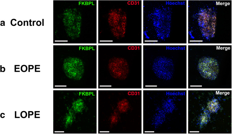
Fig. 5.

Immunofluorescent images of cardiac spheroids treated with plasma samples from healthy control, early-onset and late-onset preeclampsia patients. Cardiac spheroids were generated in hanging droplets by co-culturing primary human cardiac fibroblast cells (HCFs) and human coronary artery endothelial cells (HCAECs) in a 1:1 ratio. Following formation of 3D structures, the spheroids were treated with human plasma samples from women with or without preeclampsia. After plasma treatment, spheroids were fixated and permeabilised prior to labelling with antibodies (FKBPL, CD31) and fluorescent stain (Hoechst). a Spheroids treated with plasma from normotensive control pregnancies. b Spheroids treated early-onset preeclampsia (EOPE) patient plasma. c Spheroids treated with late-onset preeclampsia (LOPE) patient plasma
