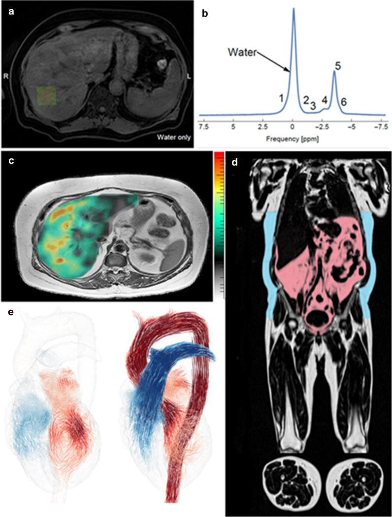
Fig. 3.
Image A shows the representative water MR image with the placement of a proton magnetic resonance spectroscopy (1H-MRS) voxel in the right hepatic lobe. Image B shows in vivo 1H-MRS spectrum for water and fat. Image C shows MRE for a cirrhotic NAFLD patient. Image E shows a whole-body water-fat separated imaging for quantification of visceral and subcutaneous adipose tissue volume. And image D shows a 4D flow image of a healthy heart. 4D flow, four-dimensional flow; 1H-MRS, proton magnetic resonance spectroscopy; MR, magnetic resonance; MRE, MR elastography

