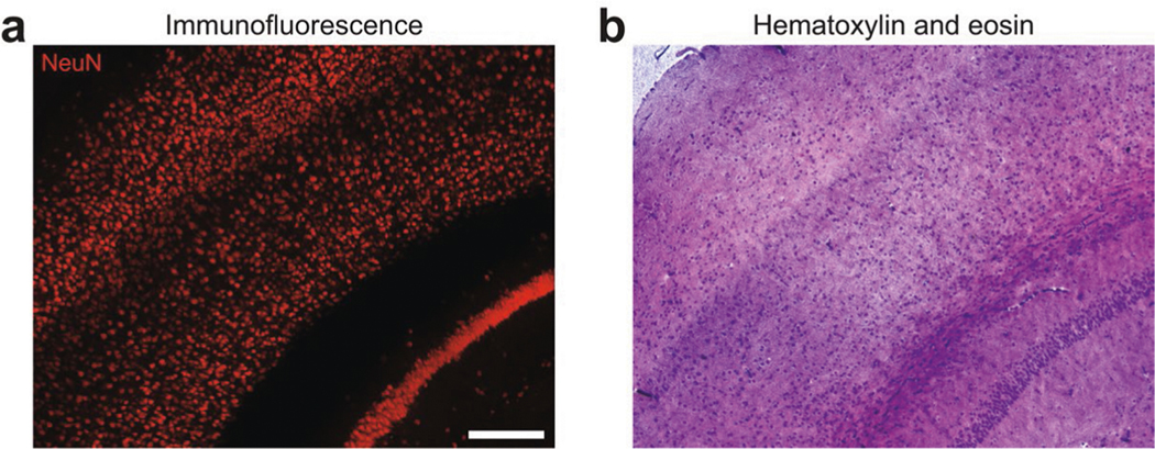
Extended Data Fig. 3 |. Downstream histology of ELASTicized tissue.
a, An ELASTicized 1-mm-thick mouse brain slice block was immunolabeled with an anti-NeuN antibody and imaged by confocal fluorescence microscopy. b, The same sample in a was then cryo-sectioned into 10-μm-thick sections. One section was histologically stained with hematoxylin and eosin with following a series of dehydration steps using ethanol solutions and xylene. The image was obtained by a bright-field microscope. Note that the image planes were not intended to match. Scale bar (adjusted to match the original tissue dimensions), 200 μm. The hematoxylin and eosin staining experiment in b was repeated six times with similar results using two NeuN-stained samples as in a.

