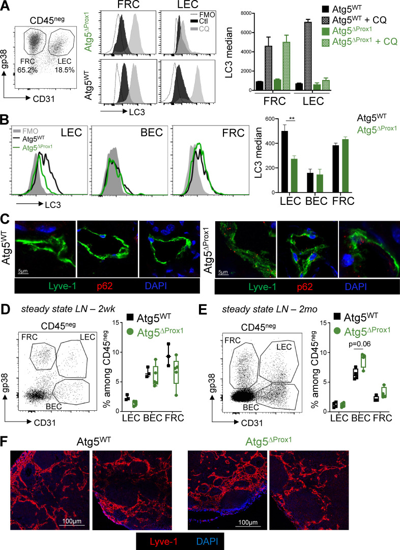
Figure 2.
Autophagy is abolished in LNs and skin LECs from Atg5ΔProx1 mice but does not impair stromal frequencies and lymphatic vessel organization in steady-state LNs. (A–F) Atg5WT and Atg5ΔProx1 mice were treated with Tx. (A) LEC/FRC cultures from LNs of Atg5WT and Atg5ΔProx1 mice were treated or not treated with CQ for 4 h, and intracellular LC3 levels (LC3-II) were assessed by flow cytometry (two independent experiments with two or three mice/group). (B) Atg5WT and Atg5ΔProx1 mice were injected with IFN-γ and CQ. CD45neg cells were isolated from skin LNs 24 h after, and intracellular LC3 levels (LC3-II) were assessed in LECs, BECs, and FRCs by flow cytometry. Unpaired t test; ** P < 0.01, three to five mice/group. (A and B) Error bars correspond to SEM. (C) Sections showing intracellular p62 in LECs (Lyve1+) from back skin of Atg5WT and Atg5ΔProx1 mice 5 d after OVA + CFA immunization. Representative images, three independent mice/group. (D and E) LEC, BEC, and FRC frequencies in skin LNs 2 wk (2wk, gated on CD45negTer119neg; D) and 2 mo (2mo, gated on CD45neg; E) after Tx treatment. Error bars correspond to lower and higher values for each group. (F) 2 mo later, lymphatic vessels (Lyve1+) were analyzed on sections of skin LNs from Atg5WT and Atg5ΔProx1 mice 2 mo after Tx treatment. Images show LNs from individual mice.

