Figure 1.
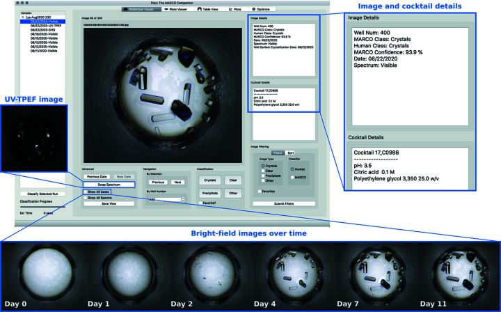
Screenshot of the Slideshow Viewer interface displaying bright-field image of Well Num 400 of a lysozyme sample set up at the Crystallization Center. Crystals are clearly visible in this well, which contains Cocktail C0988. Image Details and Cocktail Details are shown in the inset to the right. Researchers can navigate between images within the currently selected run using the Next and Previous image buttons or directly to a specific well number by typing in the well number associated with the desired image into the By Well Number box. Images can be classified via mouse clicks or keyboard shortcuts using the buttons provided under the Classification section (Crystals = 1, Clear = 2, Precipitate = 3, Other = 4). Classifying an image will automatically advance the Slideshow Viewer to the next image. Particularly interesting images can be chosen as Favorites and selected later using Image Filtering. Using the Swap Spectrum button will show UV-TPEF and SHG imaging modalities if available (UV-TPEF shown in inset to the left). Show All Dates will generate a time course of images of this particular well (shown at the bottom). Any individual image displayed by the Slideshow Viewer can also be exported as a png file using the Save View button.

