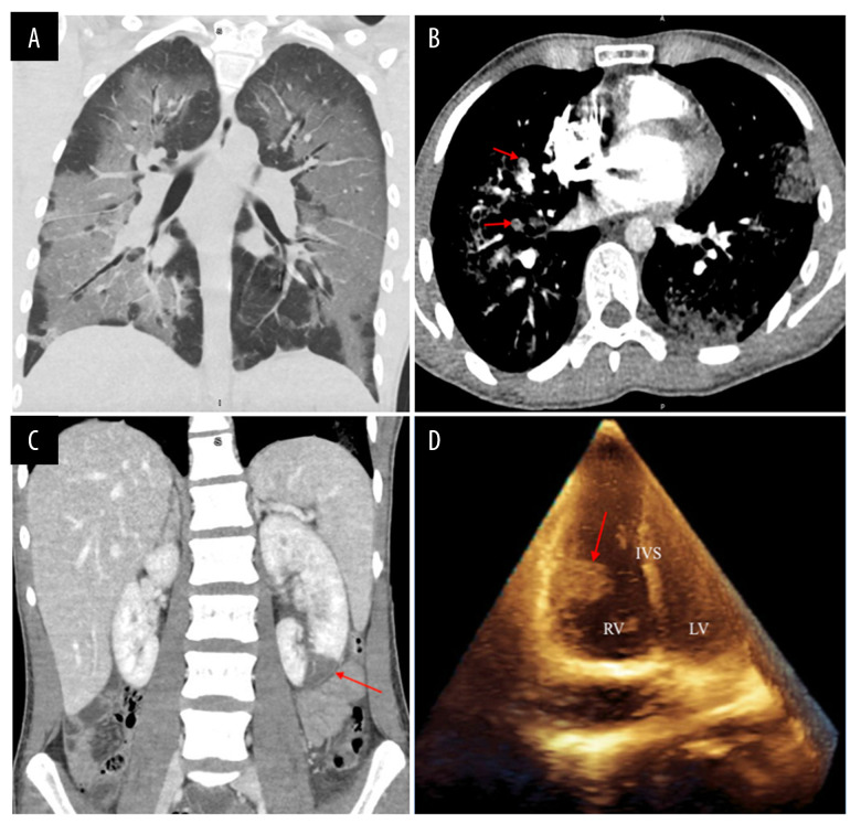
Figure 1.
(A) Computed tomography of the chest demonstrating confluent bilateral ground-glass pulmonary opacities with septal thickening and relative subpleural sparing in a lower lobe-predominant distribution; (B) Computed tomography angiography of the chest demonstrating multiple bilateral pulmonary emboli (red arrows); (C) Computed tomography of the abdomen and pelvis demonstrating a large left renal infarct (red arrows); (D) Transthoracic echocardiogram demonstrating a large mobile mass on the free wall of the right ventricle (RV – right ventricle; LV – left ventricle; IVS – interventricular septum).

