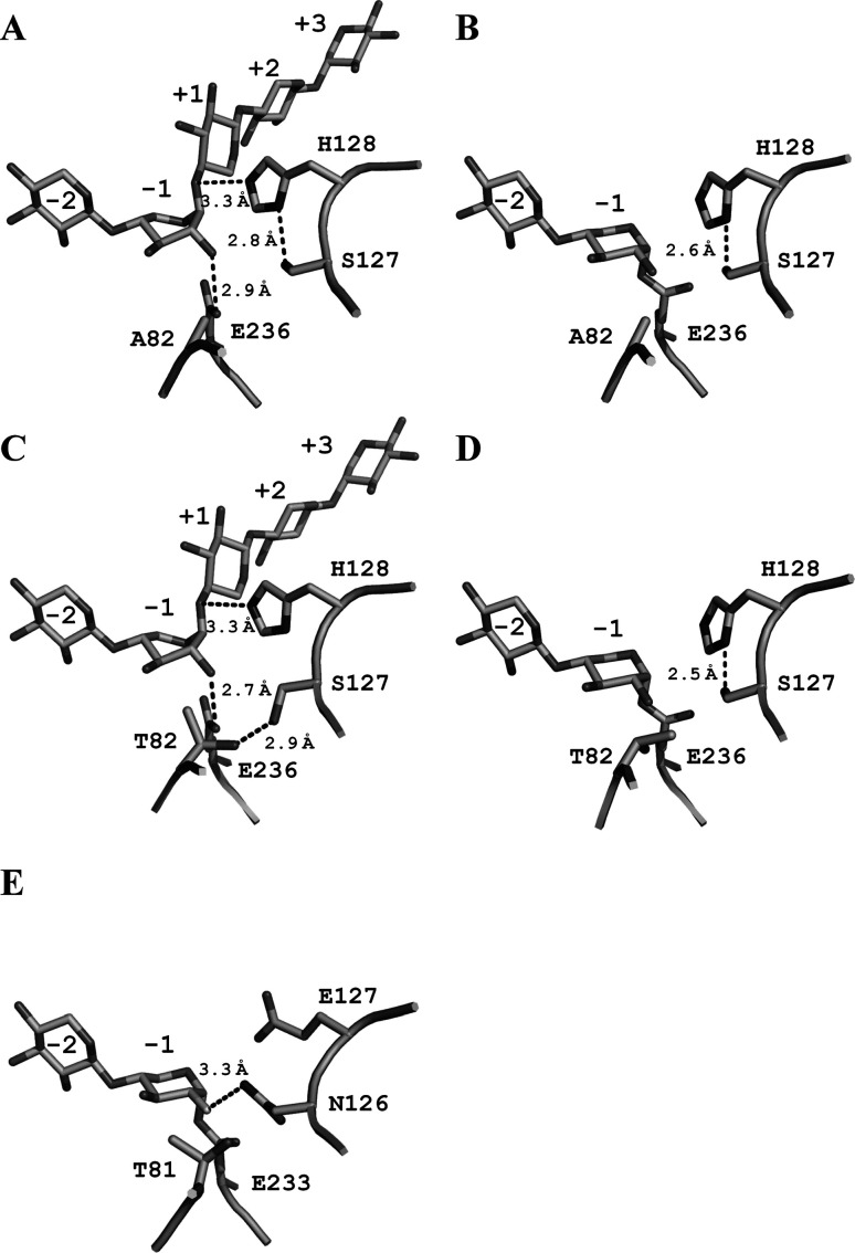
Fig. 4. Active site structures of enzyme–substrate complexes.
T82A-SEA/X5 (A), T82A-SEA/pNP-X2 (B), SEA/X5 (C), SEA/pNP-X2(E-I) (2D22) (D), and Cex in complex with 2-deoxy-2-fluoro-xylobiose (2XYL) (E). The residues essential for catalysis and bound substrates are represented by stick drawings. Hydrogen bonds are shown in broken lines. Important hydrogen-bonding distances are indicated and labeled.

