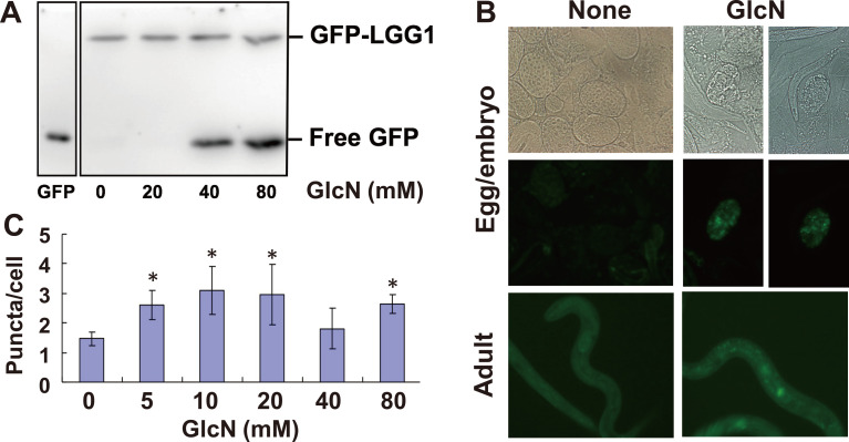
Fig. 1. Autophagy induction by GlcN in C. elegans.
(A) Transgenic worms expressing GFP::LGG-1 were grown with 0–80 mM GlcN for 96 h and harvested. GFP::LGG-1 and free GFP were detected by western blotting using anti-GFP antibody. (B) Autophagosome formation was analyzed by fluorescence microscopy. Left column, control; right column, worms grown in medium containing 40 mM GlcN. Upper row, Nomarski images of eggs/embryos; middle row, fluorescent images of eggs/embryos; bottom row, fluorescent images of adults. (C) The number of GFP-positive dots in the cytosol of seam cells was determined. Approximately 100 seam cells were analyzed. Data represent the mean ± standard deviation. Asterisks indicate significant differences compared with the control (p<0.05) by Student's t-test.

