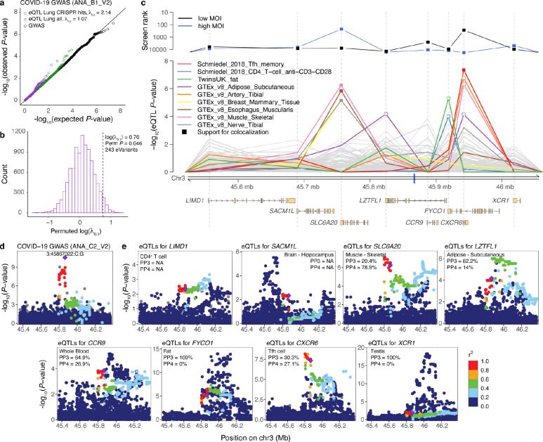
Fig. 1. Genetic regulatory effects of CRISPR hit genes and prioritization of genes in the 3p21.31 cluster associated with COVID-19 GWAS.
a, Q-Q plot showing the expected and observed P-value distribution of hospitalized vs not hospitalized COVID-19 GWAS (ANA_B1_V2) for three sets of variants: all variants tested in GWAS (black), variants that are lead eQTLs in GTEx Lung (green), and variants that are lead eQTLs in GTEx Lung for the top-ranked genes enriched in at least one CRISPR screen (purple). Inflation estimate λ0.1 measures the inflation of test statistics relative to the chi-square quantile function of 0.1. b, Histogram of the permuted log(λ0.1) to test the significance of the inflation of variants that are eQTLs for CRISPR screen hit genes (n = 243) in Lung in ANA_B1_V2 COVID-19 GWAS. c, Prioritization of genes in the 3p21.31 locus associated with COVID-19 vs population (ANA_C2_V2) GWAS. The top panel shows the ranking of the genes in the locus according to the second-best guide RNA score in the low MOI (black) and high MOI (blue) pooled CRISPR screens. The middle panel shows the eQTL P-values for the lead GWAS variant 3:45867022:C:G (denoted as a blue tick on the x-axis) in different cell types and tissues from the eQTL Catalogue and GTEx v8, 110 eQTL data sets in total. Highlighted are nine cell types/tissues, where the eQTL P-value for the lead GWAS variant is < 10−4 for at least one gene in the region. Filled square denotes suggestive support for colocalization between the GWAS and eQTL signal (posterior probability for one shared causal variant (PP4) > 0.25). The bottom panel depicts the transcripts of the eight protein-coding genes in the locus. Ranks and eQTL P-values are aligned to match the start of the gene which is shown as a grey dashed line across the panels. d-e, Regional association plots of the COVID-19 vs population (ANA-C2-V2) GWAS (d) and cis-eQTLs for the eight genes (e) from the associated locus in the cell type/tissue where the lead GWAS variant has the lowest eQTL P-value. Purple diamond denotes the lead GWAS variant, and the data points are colored based on the (weighted average) LD between the lead GWAS variant and other variants in the region in the respective study population. PP3 and PP4 - posterior probability for two different variants or one shared causal variant in coloc, respectively.

