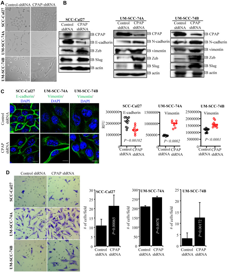
Figure 1. CPAP depleted cells show spontaneous EMT-like morphology and upregulated mesenchymal protein expression.
(A) Representative bright-field images of indicated oral cancer cell lines transduced with control-shRNA or CPAP-shRNA expressing lentiviral particles and selected using puromycin for 7 days. (B) IB showing protein levels of various EMT associated markers along with CPAP and β-actin in indicated cell lines that are stably expressing control-shRNA and CPAP-shRNA. (C) Control-shRNA and CPAP-shRNA expressing OSCC cells were fixed and permeabilized, and subjected to immunofluorescence microscopy to detect E-cadherin or vimentin (green) and nuclear stain DAPI (blue). Representative images (left panels) and relative fluorescence intensities (RFUs) of multiple cells/cell areas, quantified using ImageJ application (right panels) are shown. Scale bar: 10 μm. (D) Transwell-membrane plates with matrigel coating were used to determine the invasive properties of OSCC cell-lines. Equal numbers of control-shRNA and CPAP-shRNA expressing indicated cell-lines were seeded in serum free media in the upper chamber and incubated for 24 h. Cells on lower chamber side of the transwell membrane were stained with crystal violet, imaged and the average number (mean ± SD) of cells from at least five fields were plotted for each group. Representative results from one of the three independent experiments are shown. All P-values are by two-tailed, unpaired Mann-Whitney test.

