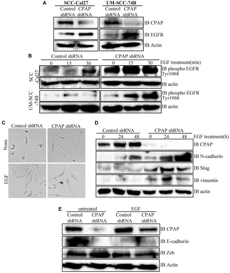
Figure 3. CPAP depletion in OSCC cells causes an increase in cellular levels of total and phospho-EGFR proteins, and EGF treatment enhances EMT-like features in these cells.
(A) Indicated OSCC cell lines that are stably expressing control shRNA or CPAP shRNA were subjected to IB to detect total EGFR and β-actin proteins. (B) These control and CPAP-depleted cell lines were maintained in serum-free media overnight, treated with cycloheximide for 1h and treated with EGF (30 ng/ml), and incubated at 37°C for indicated durations to induce signaling. Levels of phosphorylated EGFR (Tyr1068) were detected in protein equalized cell lysates by IB. (C) Representative bright-field microscopy images of control and CPAP depleted UM-SCC-74B cells left untreated or treated with EGF (30 ng/ml) for 48 h are shown. (D) IB analysis of control or CPAP-depleted UM-SCC-74B that were left untreated or treated with EGF for indicated durations and subjected to IB to detect the levels of EMT-associated proteins N-cadherin, vimentin and Slug. E-cadherin was undetectable in UM-SCC-74B cells. (E) Control and CPAP-depleted SCC-Cal27 cells were also subjected to EGF treatment for 48 h and subjected to IB to detect E-cadherin, Zeb and Slug. Vimentin and N-Cadherin were undetectable in these cells.

