Figure 3:

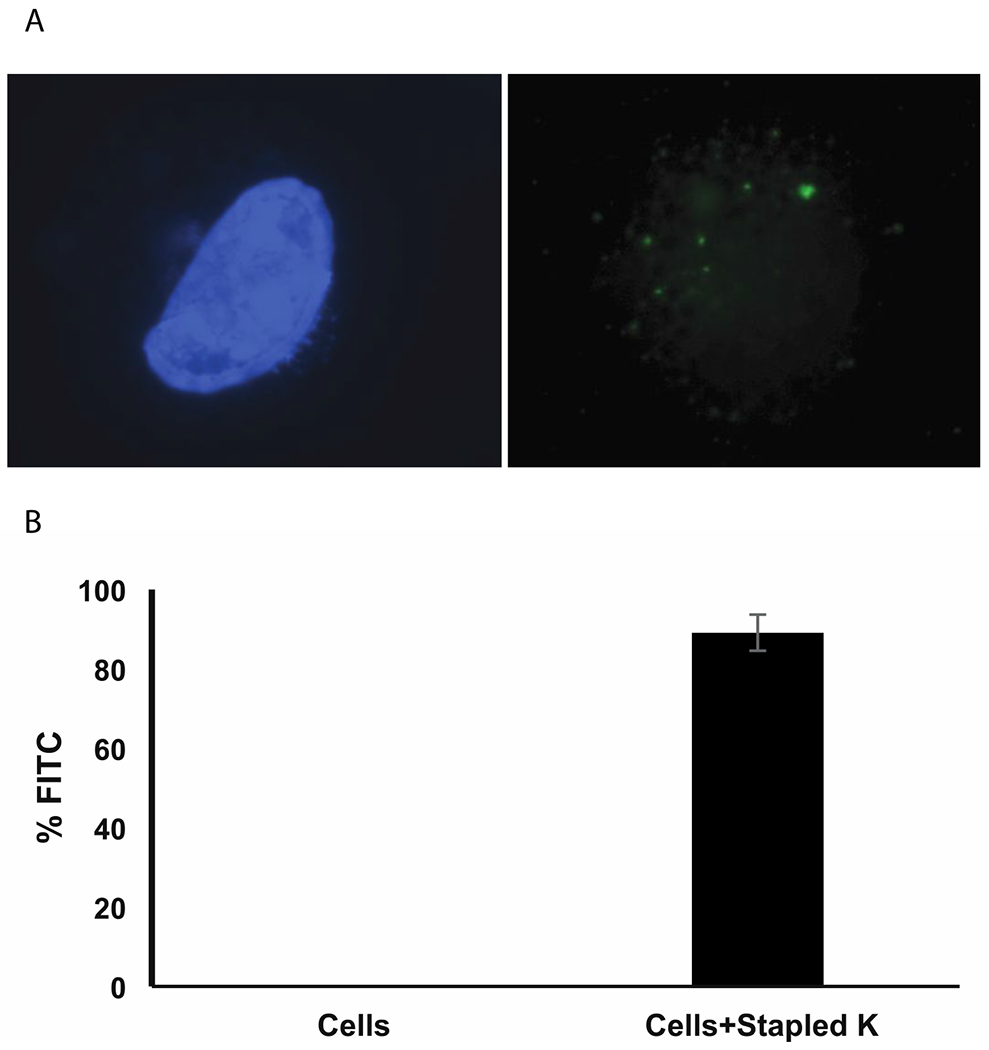
Stapled peptide K translocates into human cells in serum free media (A) Discrete foci formation of Stapled peptide K in HT1080 cells. Left panel shows blue DAPI (4’,6-diamidino-2-phenylindole) stained nuclei. Right panel shows green FITC (Fluorescein-5-isothiocyanate)-labeled stapled peptides as green foci. (B) Percentage of FITC positive HT1080 cells quantified in a flow cytometer.
