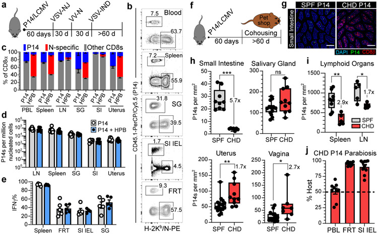
Fig. 2. The CD8+ T cell compartment expands to accommodate new and preexisting resident memory.
a-e, 60 days after LCMV, P14 immune chimeras were subjected to heterologous prime-boost (HPB) to generate a robust CD8+ T cell response against the nucleoprotein (N) epitope of VSV (a, b). Numerical abundancy, evaluated by flow cytometry (c) and QIM (d), and ex vivo functionality (e) of memory P14 CD8+ T cells in various tissues was compared and not statistically significant (p > 0.05) (d, e) between mice receiving HPB and age-matched control mice. Data shown from one of two independent experiments with similar results with n = 4-5 mice per group, per experiment (c, e). Data pooled from two independent experiments for a total of n = 8-10 mice per group (d). f-i, 60 days after LCMV, P14 immune chimeras were co-housed with pet shop mice to facilitate microbial exposure. Memory P14 CD8+ T cells in nonlymphoid (n = 7-10, 16 mice) (h) and lymphoid (n = 5,9 mice) (i) tissues were enumerated by QIM in two independent experiments (h, i). j, P14 chimeras were co-housed with pet shop mice for >60 days, conjoined via parabiosis for 30 days, at which point disequilibrium of memory P14 CD8+ T cells was assessed in n = 9-10 mice. LN, lymph node. 100 μm scale bars (g). Statistical significance was determined by two-tailed Mann-Whitney U test where *p = 0.0172, **p = 0.0028, ***p = 0.0002 (h) or *p = 0.0159, **p = 0.0040 (i). Data are presented as mean values +/− SEM or box plots showing median, IQR, and extrema.

