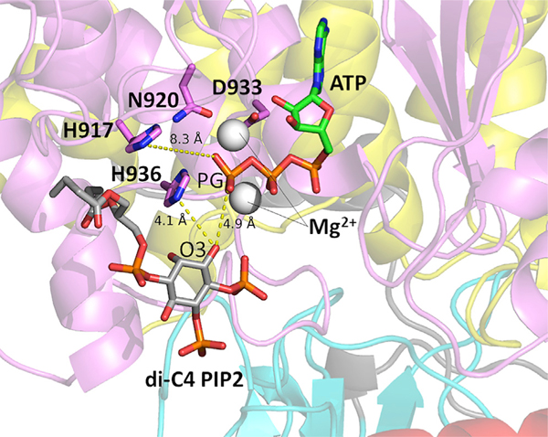
Figure 2. Putative Active Conformation of PI3Kα Obtained from Molecular Simulation.
A conformation from the simulation in which the nSH2 domain was absent is shown. Distances between the ATP PG, di-C4 PIP2, and other important active-site residues are labeled. The Nϵ H936 is only 4.1 Å away from the diC4 PIP2 O3. Mg2+ ions are represented by gray spheres.

