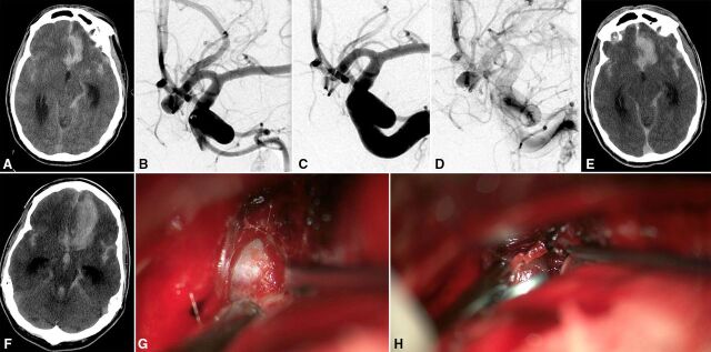
Figure 1.
Non-contrast CT on arrival to our hospital demonstrated subarachnoid hemorrhage and development of a new left frontal intraparenchymal hemorrhage, consistent with aneurysm rebleed (A). Digital subtraction angiography working view of a 6 mm anterior communicating artery aneurysm filling via a near obligate left A1 (B). Digital subtraction angiography post WEB deployment in working views (C, early arterial phase; D, late arterial phase). CT post device deployment demonstrates a stable subarachnoid hemorrhage and left frontal hematoma (E). After 4 hours, after an acute IntraCranial Pressure (ICP) spike, an interval CT scan demonstrates an enlarged frontal hematoma and new third ventricular blood (F). Intraoperative view demonstrates the WEB device in the aneurysm (G); no uncovered alternative rupture site could be identified. Postclipping intraoperative view (H).

